Acquire Your life
During your life, your brain activity can be recorded.
After the end of your flesh, your brain is mapped.
We can preserve the base of your mind.
Phase 1: Functional Recording
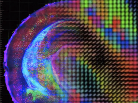
This phase is based on the old diffusion MRI (or dMRI) is a magnetic resonance imaging (MRI) method which came into existence in the mid-1980s. It allows the mapping of the diffusion process of molecules, mainly water, in biological tissues, in vivo and non-invasively.
We extended the human connectome procedure 1)into eight major domains that sample the diversity of neural activities that will be of interest to upload your mind.
For better acquirement, we suggest the use of last generation portable Noumenal reader devices. More detail on our page on noumenal technology.
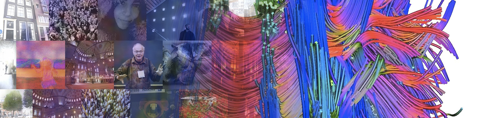
 One result of noumenal reading is a complete map of your brain connections. While you live your life, the noumenal reader gathers this map, recording also your body and environmental conditions.
This large amount of data is stored and organized in order to be ready for the next phase.
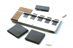 Noumenal readers are devices to recognize brain activities and incrementally record only those which are missing from the mind palimpsest they build over time.
Check out our partner PhoenixTech™ products.
Phase 2: Brain fixation
We have developed a full protocol to fixate and map your brain' structures.
It is based on the famous clarity protocol 2)- a method for chemical transformation of intact biological tissues into a hydrogel-tissue hybrid - which becomes amenable to interrogation with light and macromolecular labels while retaining fine structure and native biological molecules.
There are two main reasons for our choice:
- Connectome: it reveals local circuit wiring, relationships between neural cells, roles of subcellular structures, protein complexes, and neurotransmitters
- Storage: the fixed tissue may be stored indefinitely at 4 degC 3)
 In our clinic,your brain is sliced into more than a thousand 1cm cubes.
The entire protocol takes from 7-28 days to complete for an adult human brain, including:
- hydrogel embedding
- full lipid removal
- whole-brain antibody staining
- whole-brain high-resolution imaging (resulting in 4PB raw data)
This speed is possible because we completely automated the process and the procedure is applied in parallel for each cube slice.
Phase 3: Merging Structure and Function
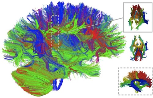
 After the acquisition of brain imaging, the data are examined in a combined model, mapping neural connectivity at the cellular level with functional tensor imaging, overcoming the limitations of classical techniques and compiling your connectome and metabolome data sets.
We use a fiber-tracking technique to join structural connectivity and functional connectivity.
With this structural-functional map, you are ready to be uploaded and become a crosser.
 All facts and characters appearing in this work are fictitious. Any resemblance to real persons, living or dead, and places is purely coincidental.